OCT – a new essential?
February 13, 2023
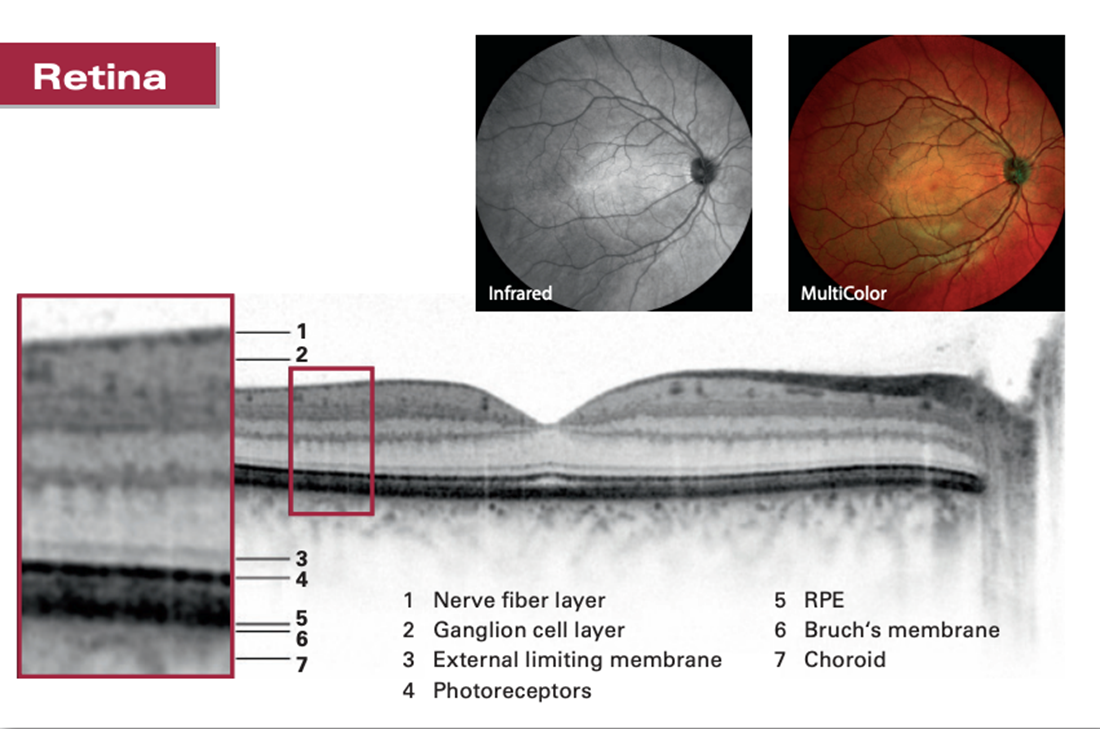
Optical coherence tomography (OCT) is a non-invasive imaging test that uses light waves to take cross sectional images of the retina, optic nerve and cornea. The high resolution means these structures can be measured in micrometres. It takes thousands of images and creates a 3D image of the structure in the eye, giving great accuracy.
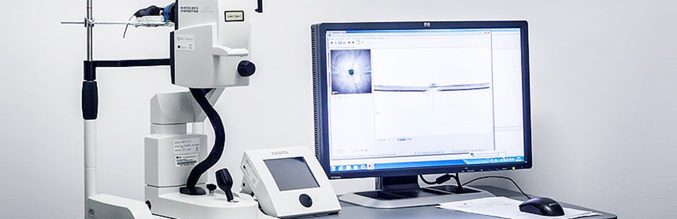
The scans are performed without touching the eye and takes only a few seconds. It can usually be performed without needing to dilate the eyes (enlarge the pupils with eye drops) and you should be fine to drive afterwards. It is not like having an MRI, for one thing, it is done sitting upright: and it also a lot less noisy!
It can pick up subtle changes to the retina and optic nerve and are particularly of use in conditions such as diabetes, age-related macular degeneration, retinal vein occlusions, macular holes and glaucoma.
The Heidelberg OCT has been taken to the international space station to look at these very structures in 2018, as astronauts can suffer from Space Flight Associated Neuro-Ocular Syndrome (SANS) and can pick up the most subtle changes that occur over time.
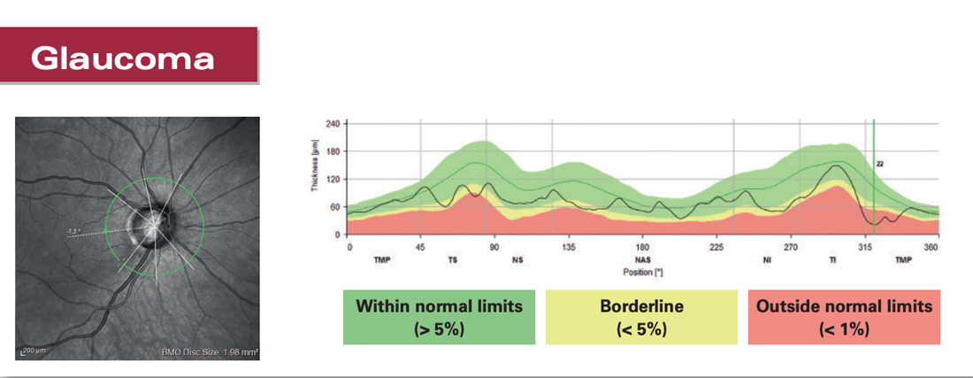
On earth, it is used to follow the progression of glaucoma. It measures cross sections of the optic nerve head and compares the thickness of the optic nerves with a database of age matched normal eyes and can do this over time. It can tell us whether a nerve is normal or not and pick up the earliest signs of glaucoma, up to four years before these become apparent to the human eye.
Although it is fast becoming an essential part of an ophthalmic exam, as with any test, the interpretation is the most important part. A change on a scan alone does not necessarily mean there is a problem. The OCT is something that is done in conjunction with taking a thorough history of the problem and examination on the slit-lamp. It is only when these three things are combined that you can make a firm diagnosis and treatment plan.